There is probably very little argument about what instrument is the neuro-ophthalmologist's best tool, viz, the ophthalmoscope. But what is the next best tool? My choice is perhaps a bit surprising and may be worth an explanation. What I have in mind is the old-fashioned manual refractometer. A modern, automatic refractometer will not do. Of course, a modern refractometer can estimate the optical parameters in tricky refraction situations, but among all ophthalmic instruments it is only the manual refractometer that can illuminate the eye's optical quality. The explanation for this remarkable ability is that the manual refractometer uses a double-pass of image-forming rays through the eye under examination. The instrument sums the effects of any and all optical imperfections twice, once as the rays head towards the retina, and then again as they exit the eye. This double summing makes it very easy for the examiner to recognize the operation of multiple, minor optical imperfections. Taken one by one, small optical imperfections may appear negligible but they may sum up in a highly significant manner. Hence, the simple act of estimating image quality in a manual refractometer may help to unravel the cause of otherwise unexplained visual loss.
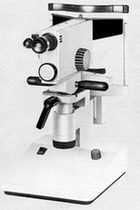
The instrument I use is an old Rodenstock PR50, salvaged from the scrapheap when the department decided to switch to automatic refractometers, throwing out the child with the bathwater. The Zeiss company has produced a similar instrument. There may be others, too.
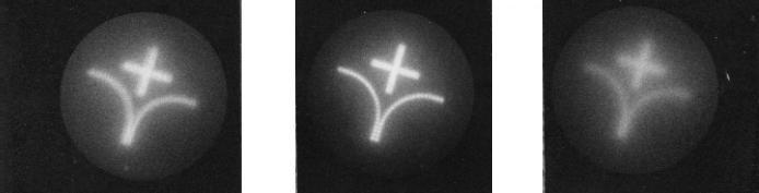
Looking into the PR50 ocular, the image of a classical cross-and-arrow target can be seen, reflected off the fundus. After focusing, the image is scrutinized with regard to sharpness and brightness. The sharpness reflects the imaging quality of the eye under examination, of course. It is easy to learn to predict visual acuity from the level of sharpness. It is useful to assess image brightness as well. The brightness deteriorates rapidly with increasing amounts of optical opacity, e g, from nuclear sclerosis. This seems to be another aspect of visual imapirment that is very difficult to capture by other tools.
The PR50 was not designed for objective recordings of image quality but it can be tweaked, with considerable effort, to allow photograhy. The center image below illustrates image quality in a normal eye whereas the flanking images were obtained from eyes with fairly subtle nuclear sclerosis. Higher degrees of opacity could not be recorded because of the associated reduction of image brightness: the images shown below required the film to be pressed to no less than 3200 ASA.
Optical double-pass refractometers don't seem to be manufactured nowadays.
Digital double-pass instruments are just beginning to appear. They should have a similar potential to aid identification of optical causes of unexplained visual loss.

 The instrument I use is an old Rodenstock PR50, salvaged from the scrapheap when the department decided to switch to automatic refractometers, throwing out the child with the bathwater. The Zeiss company has produced a similar instrument. There may be others, too.