Rarebits or briefly flashed receptive field-size test targets were introduced in 2002
[1]
in an attempt to resolve the many shortcomings of traditional ways of testing vision (see
[A,
B,
C]
for on-site overviews). Rarebit tests essentially look for holes in the neuro-retinal receptive field matrix. First out
were RareBit Perimetry (RBP) and the RareBit Fovea Test (RFT)
[D,
1],
which were followed by a self-contained macula test, MacuBit
[E,
2].
Several independent reports have attested to the utility of the rarebit concept
[F].
One of the novel features of the original rarebit tests was the simulta- neous presentation of two widely spaced test targets and asking the subject to indicate whether he or she saw two light dots, one, or none at all, by clicking twice, once, or not at all, on the computer mouse. The paired target feature not only saved on test time but it also provided a means for checking for false responses. Unfortunately, some very young and also some elderly subjects found mouse-clicking difficult, so an alternative approach was sought. When considering the option of having subjects verbally calling out their perceptions (with the examiner recording their responses), it comes natural to consider presentations employing more than two rarebits. Such "multibit" presentations might employ easily recognized geometric or alphanumeric symbols, constructed in such a way that failure to perceive one or more of the component rarebits would preclude correct identification. Further, to estimate the severity of any neural matrix defects, the number of component rarebits could be increased until identification is achieved.
Interestingly, multibit test targets can be made to subtend several degrees of visual angle, i e, much more than, say, a 0.1 (20/200) optotype (0.8°), or the largest Goldmann perimeter target (1.7°), to quickly probe large areas of the neural matrix. Very large targets cannot cannot provide detailed maps, of course, so such targets may be best suited for quick overviews. It may be argued that quick overviews are all that is needed in clinical work, provided that their rationales are sound.
Examples of multibit test targets
Swarms It is an everday experience that the approximate number of members in a group can be estimated at a glance. One way of exploiting this capacity is to present, say, 10 rarebits, randomly distributed within a circumscribed area subtending, say, 5°. The test task is to decide whether the number of rarebits seen actually equals 10. This would not be the case in the presence of neural matrix damage: some rarebits would then fall into matrix holes and so escape detection. In such cases, the number of rarebits shown would need to be increased to reach the criterion number. Informal explorations have attested to the feasibility of the swarm estimation approach but they have also highlighted an insufficient precision.
Clock faces have the advantage of instant familiarity. They also have the useful and perhaps unexpected property of escaping cerebral filling-in. Intentional removal of one or more tick marks is easily recognized. On the other hand, it is difficult to arrange for complete percepts in the presence of matrix defects. Therefore, clock face targets seem useful for pass-fail screening only, not for quantitative estimations.
Segmented digits also have the advantage of instant familiarity, thanks to the everywhere presence of digital displays. Other advantages include the predictability of the minimum number of rarebits normally required for recognition, namely, 3 rarebits per segment, and the ease of increasing this number if recognition fails. To keep the number of rarebits reasonably constant across different digits, 1 and 7 have to be excluded. Further, in the basic configuration, 8 differs from 0 with respect to one rarebit only, so it appears sound to exclude 8, too. Hence, digits 0, 2, 3, 4, 5, 6, and 9 should be most useful.
Segmented letters selected along similar lines should be equally effective.
First implementations
Several variations on the multibit theme have been explored:
The FlashField test is a qualitative pass-fail adaptation of the set-up used in rarebit perimetry
[D],
where the original rarebit pairs are replaced by multibit clock faces centered on the fixation point. There are 5 clock faces, with radii subtending 5 to 22°. The examiner selects a radius and flashes a presentation of 200 ms duration. The subject's task is to call out any apparently missing tick marks. The quality of performance can be checked by the deliberate removal of tick marks. This test is presently being evaluated by independent investigators. Continuously running variants are currently being explored.
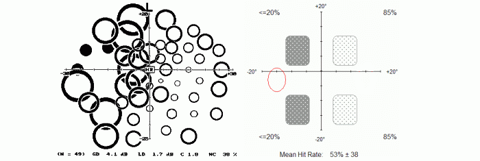
The DigitField test was devised to try very large (8x10°) segmented digits as a rapid quantitative visual field test, again in the rarebit perimetry set-up. This approach appears useful inside the eccentricity of the blind spot only: it is difficult to read multibit digits more peripherally. Shown below are left-eye results from a subject with a mid-chiasmal lesion, using high-pass resolution perimetry
[G]
(left) and the DigitField test (right). The different number of target locations (50 versus 4) is reflected in the test durations (ca. 5 and l.5 minutes, respectively). It is interesting to ponder whether the longer and more detailed examination provides grounds for a better clinical evaluation.
The MacuDigBit test uses segmented digits in the MacuBit set-up
[E].
This is a quantitative test where the examiner controls the number of multibits making up the test digits. Initial trials have proved the feasibility of the concept. The main advantages over the original implementation are that subjects don't need to master mouse-clicking and that test durations are approximately halved. The main disadvantage is that results cannot be given on a finely graded scale.
The DigitScreen test is an intuitive, super-fast screener of the central-most visual field. It runs inside this browser and may merit a more detailed presentation:
DigitScreen: a fast multibit screening test
This test presents pairs of randomly selected segmented digits in random screen locations. The pair members are shown with a slight delay in-between, as exemplified to the right. This arrangement makes for a simple test task and a large target area. The test cycles
continuously over 3 test levels. These levels employ 3, 4, and 5 rarebits per digit segment, respectively. Each cycle presents a total of 95 rarebits, on average. The cycle duration is ca. 8 seconds.
The subject's task is simply to call out any seen digits. There are no fixation demands.
The examiner's task is simply to count the number of correctly read digits for each cycle of 3 presentations. Many normal subjects need no more than one cycle to complete the test. Other subjects may need a few cycles to return consistent results. In case of variation it is sound to take the average of, say, 3 cycles.
Normal subjects should read 5 or 6 digits correctly in each cycle, depending on age. The first results from normal subjects suggest that 6 digits should be expected from those younger than approximately 60 years whereas those above 60 should read at least 5. 4 digits, or less, are abnormal, with smaller numbers representing more severe damage.
The number of digits read is a summary index of the state of the central part of the neuro-retinal matrix. A summary index obviously cannot provide any topographic information. However, some topographic information can be gained during the test by paying attention to the locations of any missed digits. For example, a patient with a right homonymous field defect will generally read the left-hand pair members more easily than the right-hand ones.
Use a stand-alone liquid-crystal display (LCD) with a diagonal of ≥15" and a vertical resolution of ≥800 pixels. The test uses the central 1024 x 768 pixels only. Laptop displays are not recommended because of their lower contrast. Use the screen manufacturer's default settings for resolution, brightness and contrast. The browser borders and any display indicator lights need to be masked to prevent disturbing stray light. The most practical solution may be to lean a cardboard mask against the front of the display. The test space should be completely dark. Arrange for comfortable viewing at 4 m distance. Cover one eye. Use a proper ametropia correction.
The test area subtends ca. 4.5 x 3.5° at 4 m test distance, the test digits subtend ca. 0.6 x 0.8° (= 0.1 decimal, 20/200 Snellen), and the multibits ca. 0.5', assuming a pixel size of ca. 0.3 mm. Presentation time is 150 milliseconds.
DigitScreen needs a 4-meter test distance. Paradoxically, the test becomes increasingly difficult with shorter distances and is almost undoable at a reading distance. The perceptual capacity to group briefly exposed dots into patterns is apparently inversely related to the pattern's angular subtense.
A first study of DigitScreen's diagnostic capacity involved 30 normal subjects and 47 patients with various optic nerve or visual pathway lesions of light to moderate severities. DigitScreen's power of discrimination was compared to those of acuity and high-pass resolution perimetry by means of receiver-operating characteristics. The areas under the curves equalled 0.87, 0.77, and 0.78, respectively. DigitScreen clearly ranked highest but the advantage over the runner-ups fell just short of statistical significance (p = 0.07). Sensitivities were estimated to 67%, 48% and 54%, respectively. It may be concluded that DigitScreen test has a remarkable diagnostic potential, in spite of its super-short test duration. See
[3]
for full details.
 One of the novel features of the original rarebit tests was the simulta-
 Swarms It is an everday experience that the approximate number of members in a group can be estimated at a glance. One way of exploiting this capacity is to present, say, 10 rarebits, randomly distributed within a circumscribed area subtending, say, 5°. The test task is to decide whether the number of rarebits seen actually equals 10. This would not be the case in the presence of neural matrix damage: some rarebits would then fall into matrix holes and so escape detection. In such cases, the number of rarebits shown would need to be increased to reach the criterion number. Informal explorations have attested to the feasibility of the swarm estimation approach but they have also highlighted an insufficient precision.
 Clock faces have the advantage of instant familiarity. They also have the useful and perhaps unexpected property of escaping cerebral filling-in. Intentional removal of one or more tick marks is easily recognized. On the other hand, it is difficult to arrange for complete percepts in the presence of matrix defects. Therefore, clock face targets seem useful for pass-fail screening only, not for quantitative estimations.
 Segmented digits also have the advantage of instant familiarity, thanks to the everywhere presence of digital displays. Other advantages include the predictability of the minimum number of rarebits normally required for recognition, namely, 3 rarebits per segment, and the ease of increasing this number if recognition fails. To keep the number of rarebits reasonably constant across different digits, 1 and 7 have to be excluded. Further, in the basic configuration, 8 differs from 0 with respect to one rarebit only, so it appears sound to exclude 8, too. Hence, digits 0, 2, 3, 4, 5, 6, and 9 should be most useful.
 This test presents pairs of randomly selected segmented digits in random screen locations. The pair members are shown with a slight delay in-between, as exemplified to the right. This arrangement makes for a simple test task and a large target area. The test cycles
continuously over 3 test levels. These levels employ 3, 4, and 5 rarebits per digit segment, respectively. Each cycle presents a total of 95 rarebits, on average. The cycle duration is ca. 8 seconds.
This test presents pairs of randomly selected segmented digits in random screen locations. The pair members are shown with a slight delay in-between, as exemplified to the right. This arrangement makes for a simple test task and a large target area. The test cycles
continuously over 3 test levels. These levels employ 3, 4, and 5 rarebits per digit segment, respectively. Each cycle presents a total of 95 rarebits, on average. The cycle duration is ca. 8 seconds.