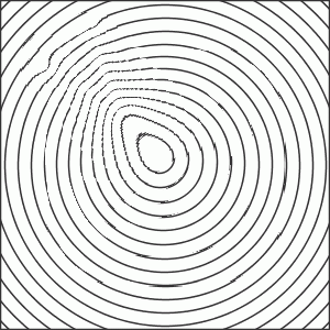
It is an everyday experience that there is an inverse relation between size and contrast: the smaller the target, the greater the contrast needed for visualization. The image to the right illuminates the curvilinear relationship, the contrast sensitivity curve (the border zone between seen and not seen parts of the bars).
The image represents size (or more precisely, spatial frequency) along the horizontal axis whereas contrast is varied vertically. (The image contrast is most likely somewhat inaccurate, because of limitations of the current medium. Visit the source, the
Ohzawa Laboratory,
for better renditions.) It is interesting to try various viewing distances to see how the curve changes its shape.
Contrast sensitivity tests aim to illuminate visual performance at contrast levels lower than those of acuity tests. Contrast sensitivity tests have not made major inroads in clinical work. One reason is that early hopes of identifying visual loss restricted to low or intermediate spatial frequencies appear to have been frustrated (high frequencies are exploited by acuity tests) . Another reason is the difficulty of calibration. Further, most tests are fairly difficult. The reproduction of the popular
Pelli-Robson
chart (right) is most likely inaccurate as to contrast levels but it may still serve to illustrate the effort required to find and decipher letters at near-threshold contrast. Note that all the letters have the same size. The test distance is also fixed, at 1 m. Hence, the Pelli-Robson chart assesses the vertical height of the contrast sensitivity curve in a restricted band across the horizontal spatial frequency axis.
Tri-VA: a one-minute test procedure
One way to simplify the test task is to traverse the contrast sensitivity gamut horizontally instead of vertically (as done by the Pelli-Robson test), by using targets with a fixed, sub-maximal contrast level and estimating the smallest size that can be seen. This is the approach of the Tri-VA display below, which uses so-called high-pass, spatial frequency filtered targets of low contrast. Targets of this type have a peculiar, "vanishing" property in that they are either resolvable or invisible, which makes for a simple test task. Vanishing targets have proved very useful in another setting, namely,
high-pass resolution perimetry.
Tri-VA uses a test pattern made up of three bar-shaped targets of different sizes. The bars are arranged like spokes or prongs that radiate from a central dot of high contrast. The dot serves to aid localization. Each prong has a bright core and a dark rim. Prong dimensions and luminances are balanced in such a way that the space-average luminance equals that of the background. The test should be run in a dark room. The suggested test distance is 3 m
The subject's task is simply to count the number of prongs seen as the size of the pattern is varied. Right-click on the test display to increase the pattern size and left-click to decrease the pattern size. The goal is to find that pattern where the subject can see one or two prongs only. If three prongs are seen, pattern size needs to be decreased and if none is seen, pattern size needs to be increased. If the response is two prongs seen, it is the mid-size prong that represents the threshold. If one prong only is seen, its size represents the threshold. The subject should be asked to look quite carefully before responding.
Current prong core widths are shown at the bottom of the display. When the smallest visible prong has been found, make a note of its core width: this is the test result.
Calibration.
The display offers one adjustment only, namely, the luminance of the dark prong rims. Adjustment is best made at a 3 m viewing distance, with the aid of an assistant. Begin by finding what prong size is closest to threshold at the selected distance. Then increase the viewing distance slightly, to the point where the prong under consideration just begins to disappear from view. Vary the viewing distance slightly and judge the overall impression of the prong just as it melts into the background. If the prong appears darker than the background, the assistant should increase the rim luminance setting by tapping the Increase button one or more times. If the prong appears lighter than the background, the assistant should decrease the rim luminance setting by tapping the Decrease button. Calibration is finished when the prong invisibly melts into the background, without passing through a darker or brighter stage. Record the luminance setting for future reference. Exact definition of the contrast level requires fairly sophisticated measuring equipment but is not really necessary as long as testing is performed under constant conditions, with one and the same computer set-up. Normative values have to be defined individually for each set-up, from test results obtained from normal subjects. The luminance values are nominal only and do not necessarily belong to a linear scale.
This test was originally developed in C under the name Y-VA in 1994, as part of the DOS-based
Ophthimus System.
Both Y-VA and Tri-VA are known to be well-behaved
[1,
2,
3].
How an abnormal contrast sensitivity relates to the state of the neural matrix is currently poorly known. Likewise, it is poorly known what role contrast sensitivity testing may have in critical testing of central vision.
Dysmetropsia
Dysmetropsia means seeing that objects have unaccustomed dimensions. Micropsia is more common than macropsia. The two may be mixed, often in a spatially irregular fashion, to produce metamorphopsia. Static and dynamic representations can be viewed
elsewhere
on this site. The most common variants are due to macular diseases that cause deformations and dislocations within the retinal neural matrix. If bilateral, retinopathic dysmetropsias are rarely perfectly symmetrical. A dysmetropsia that is perfectly symmetrical between the eyes is more rarely encountered. It is usually transient and often localized to homonymous hemifields. This latter variant is usually attributable to cortical dysfunction, often in a migraine-related setting.
The most common test for dysmetropsia is the Amsler chart, a square grid of black lines on a white background
[4].
The test task is to examine the grid for any irregularities while steadily fixating on a central dot. Any irregularities can be sketched on the chart to provide a permanent record for future reference. Several variants involving color and contrast have been suggested during the years but the original chart seems to reign supreme. Yet, there are several drawbacks. For one thing,
dysmetropsic loci may hide in-between the grid lines, and for another, retinopathies are often associated with rampant
Troxler
fade-from-view effects, making for a difficult test task. A dynamic display should facilitate detection of localized dysmetropsia.
The MetaScreen display presented in the left panel below uses flowing circles rather than squares for a more uniform appearance. The symmetrical flow should enhance fixation stability at the same time as it seamlessly sweeps the test area and counteracts the Troxler effect. Click on the display to start movement, click again to stop. There are no adjustable parameters. The test task is to identify any localized deviations from true circular shapes, while watching the display's center. The right panel shows a simulated snapshot of what a localized dysmetropia might look like.
The MetaScreen display induces a powerful counter movement upon stopping the flow. This is an example of the
waterfall illusion.
An important disadvantage of qualitative dysmetropsia testing is that it is impossible to know how good an observer the patient is. The dynamic MetaFlow test (formerly named MacuFlow)
[5]
was devised to correct this deficit and to provide a quantitative record. The idea is to attempt nulling of any dysmetropsia by actively deforming the grid in the opposite direction.
The MetaFlow test sweeps concentric square or octagonal outlines across the test area. The flowing outlines can be deformed via the keyboard. This active deformation feature allows testing of the patient's powers of observation and allows an approximate correction of any dysmetropsia. The display subtends approximately 20° at 0.3 m viewing distance. To avoid clutter, the test opens in a separate window.
Click on the display to start the flow, click again to stop. Tap the keypad keys to contract the outlines in the direction of the tapped key. For example, tap key 1 to contract the pattern in the lower left octant. To make the pattern expand instead, hold down the shift key and tap key 1. Tap the Delete key to restore the regular pattern. When done, tap the s key to obtain a statistical representation of deformations, including mean unsigned deviation ± standard deviation. Tap the s key again to hide the numbers.
Various settings are accessible under the Settings menu. Shape toggles between square and octagonal outlines, Kbd step sets the magnitude of change produced when tapping the keypad keys whereas Bias allows setting emphasis on central or peripheral parts of the pattern. The remaining functions should be self-explanatory.
Amsler-type tests are often used to assist in the detection of scotomata. The MetaFlow test provides the opportunity to "punch out" a part of the pattern. This feature may help to define the patient's power of observation. The punch size can be selected under the Punch menu. Each click selects a new location.
Test results can be stored as image files. A simple way is to tap the PrintScreen keyboard key, open MSPaint, and select Edit/Paste. The image can then be cropped as desired and saved in a variety of formats.
By design, the MetaFlow Test attempts to null spatially extensive dysmetropias. For localized defects, the MetaStat Test may prove more useful. To facilitate localized attention, the test pattern - a square grid - remains stationary. As mentioned above, small localized defects may hide in-between grid lines. The MetaStat test minimizes the risk of missing such defects by offering three different grids. The coarse, medium and fine meshes aim to assess gross, intermediate and subtle metamorphopsias, respectively. Optionally, the grid can be rendered like a chessboard.
The test field subtends approximately 13° at 0.3 m viewing distance. It is smaller than that of the MetaFlow test to allow larger deformations.
The display opens with a localized deformation. Point out the deformed area and show how to move the drag dot into this area, by holding down the Ctrl keyboard key while tapping one of the Arrow keys. Then release the Ctrl key and use the Arrow keys to drag the selected intersection into the correct position. Repeat until the subject masters the procedure. Tap the Del key to restore the regular pattern.
Select a suitable mesh size under the Settings menu. Ask whether the subject perceives any distortion while fixating the central ring target. If so, move the drag dot into the deformed area and try to correct the apparent deformity. When done, tap the s key to obtain a statistical estimate of the compensatory deformation, in the form of the mean unsigned deviation ± standard deviation. Tap the s key again to hide the numbers.
To obtain an assessment of the subject's power of observation, introduce a deliberate deformation within a subjectively normal area of the test field and ask for correction. Any deviations from full correction illuminate the power of observation.
Test results can also be stored as image files. A simple way is to tap the PrintScreen keyboard key, open MSPaint, and select Edit/Paste. The image can then be cropped as desired and saved in a variety of formats.
Various settings are accessible under the Settings menu. Kbd step sets the magnitude of change produced when tapping the keypad keys. The remaining functions should be self-explanatory.
Because the MetaFlow and MetaStat tests cover fairly extended visual field areas, they are not useful for revealing minimal central-field dysmetropsia as a cause of minimal vision loss. A simple subjective comparison of apparent sizes and shapes between the eyes, for example, when looking at an acuity chart, may be more helpful in this setting. It is best to direct the subject to judge an optotype size that is far above the resolution threshold, say, twice as large, to facilitate concentration on the comparison of size and shape. Also, it may be helpful to direct the patient's attention to one specific letter, e g, in the middle of the selected line. The examiner should apply alternate cover to aid evalution.
Mapping macular scotomata - the GridBlock Test
The Amsler grid is often used to search for central/paracentral scotomata [
4].
Then, the test task is to fixate the grid center and to identify any portions of the grid that appear to be missing or poorly defined and to mark such areas on the grid. These are difficult tasks, not the least because of the operation of Troxler fade-from-view and cerebral filling-in processes. It is not possible to assess the quality of the subject's performance. Clearly, automated perimetry with a "macula grid" would be preferable but may be difficult to arrange with the frequency needed for careful monitoring of critical conditions like neo-vascular macular disorders. Hence, there is a need for a self-administered test that circumvents the limitations of the Amsler grid. The GridBlock Test was devised to meet this need
[5].
The idea behind the GridBlock Test is to break down the 20x20° Amsler grid into 2x2° blocks. The blocks are rendered with interrupted lines to minimize redundant information. Optionally, the blocks can be rendered with dots, similar to rarebit swarms. Blocks are shown one by one, in random order, interspersed with control presentations. Exposure time is 200 ms, i e, short enough to frustrate changes of fixation and to prevent Troxler effects.
There are 88 complete blocks that seamlessly tile the display area, except for its corners, and 12 incomplete blocks that serve as controls, for a total of 100 presentations. Examples of complete (left) and incomplete blocks (right) are shown in the figure. The current presentation number is shown in the display center, with the aim to encourage stable fixation and to provide an indication of how far the test has progressed. The test task is to click the mouse button when and only when a complete block is seen. A response to a control presentation (an incomplete block) causes negative feedback in the form of a red "traffic light" and a break in the test rhythm.
At the end of the test, all blocks seen as complete will be shown on the display together with error statistics that illuminate the subject's performance. Testing time is about 2.5 minutes using the default settings. Recommended viewing distance is 0.3 m.
Right-click on the display to start; right-click again to take a pause. Left-clicks are reserved for responses. The mouse cursor should be kept in the test area's lower left or right corner during the test. The rate of presentations can be selected under the Settings menu. The Pattern menu allows a choice between blocks defined by line segments and dots, respectively, and offers the option of inverting contrasts.
There are three types of control presentations: complete blanks, half blocks, and three-quarter blocks. There are four controls of each type. Controls are selected and located at random. Any responses are counted separately for each type and presented in a small/medium/large sequence at the end of the test. For example, 50/25/0 means that the subject responded to 50% of blocks lacking one quadrant, to 25% of half blocks, and to none of the full blanks.
Test results can be stored as image files. A simple way is to tap the PrintScreen keyboard key, open MSPaint, and select Edit/Paste. The image can then be cropped as desired and saved in a variety of formats.
 It is an everyday experience that there is an inverse relation between size and contrast: the smaller the target, the greater the contrast needed for visualization. The image to the right illuminates the curvilinear relationship, the contrast sensitivity curve (the border zone between seen and not seen parts of the bars).
The image represents size (or more precisely, spatial frequency) along the horizontal axis whereas contrast is varied vertically. (The image contrast is most likely somewhat inaccurate, because of limitations of the current medium. Visit the source, the
Ohzawa Laboratory,
for better renditions.) It is interesting to try various viewing distances to see how the curve changes its shape.
It is an everyday experience that there is an inverse relation between size and contrast: the smaller the target, the greater the contrast needed for visualization. The image to the right illuminates the curvilinear relationship, the contrast sensitivity curve (the border zone between seen and not seen parts of the bars).
The image represents size (or more precisely, spatial frequency) along the horizontal axis whereas contrast is varied vertically. (The image contrast is most likely somewhat inaccurate, because of limitations of the current medium. Visit the source, the
Ohzawa Laboratory,
for better renditions.) It is interesting to try various viewing distances to see how the curve changes its shape.
 Contrast sensitivity tests aim to illuminate visual performance at contrast levels lower than those of acuity tests. Contrast sensitivity tests have not made major inroads in clinical work. One reason is that early hopes of identifying visual loss restricted to low or intermediate spatial frequencies appear to have been frustrated (high frequencies are exploited by acuity tests) . Another reason is the difficulty of calibration. Further, most tests are fairly difficult. The reproduction of the popular
 Tri-VA uses a test pattern made up of three bar-shaped targets of different sizes. The bars are arranged like spokes or prongs that radiate from a central dot of high contrast. The dot serves to aid localization. Each prong has a bright core and a dark rim. Prong dimensions and luminances are balanced in such a way that the space-average luminance equals that of the background. The test should be run in a dark room. The suggested test distance is 3 m
. Source: www.iris-ward.com") The most common test for dysmetropsia is the Amsler chart, a square grid of black lines on a white background
[
The most common test for dysmetropsia is the Amsler chart, a square grid of black lines on a white background
[
 The MetaFlow test sweeps concentric square or octagonal outlines across the test area. The flowing outlines can be deformed via the keyboard. This active deformation feature allows testing of the patient's powers of observation and allows an approximate correction of any dysmetropsia. The display subtends approximately 20° at 0.3 m viewing distance. To avoid clutter, the test opens in a separate window.
 By design, the MetaFlow Test attempts to null spatially extensive dysmetropias. For localized defects, the MetaStat Test may prove more useful. To facilitate localized attention, the test pattern - a square grid - remains stationary. As mentioned above, small localized defects may hide in-between grid lines. The MetaStat test minimizes the risk of missing such defects by offering three different grids. The coarse, medium and fine meshes aim to assess gross, intermediate and subtle metamorphopsias, respectively. Optionally, the grid can be rendered like a chessboard.
The test field subtends approximately 13° at 0.3 m viewing distance. It is smaller than that of the MetaFlow test to allow larger deformations.
By design, the MetaFlow Test attempts to null spatially extensive dysmetropias. For localized defects, the MetaStat Test may prove more useful. To facilitate localized attention, the test pattern - a square grid - remains stationary. As mentioned above, small localized defects may hide in-between grid lines. The MetaStat test minimizes the risk of missing such defects by offering three different grids. The coarse, medium and fine meshes aim to assess gross, intermediate and subtle metamorphopsias, respectively. Optionally, the grid can be rendered like a chessboard.
The test field subtends approximately 13° at 0.3 m viewing distance. It is smaller than that of the MetaFlow test to allow larger deformations.
 The idea behind the GridBlock Test is to break down the 20x20° Amsler grid into 2x2° blocks. The blocks are rendered with interrupted lines to minimize redundant information. Optionally, the blocks can be rendered with dots, similar to rarebit swarms. Blocks are shown one by one, in random order, interspersed with control presentations. Exposure time is 200 ms, i e, short enough to frustrate changes of fixation and to prevent Troxler effects.